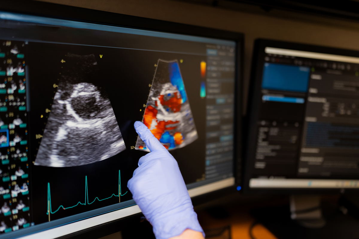
From early disease detection to workflow relief, echocardiography’s evolution is giving clinicians sharper insight – and patients clearer answers.
Over the past decade, echocardiology has quietly transformed diagnostic confidence in cardiovascular care – often in ways clinicians may not fully recognize. As Jacob Dewees, Clinical Luminary Leader, Cardiovascular Ultrasound at GE Healthcare, explained, “the very first line of visual interpretation of the heart” comes through echo, long before other modalities can reveal how the heart is actually functioning.
That accessibility has helped fuel rapid innovation. “The one beautiful thing about echo is that it’s really easily accessible,” Dewees said, noting that ultrasound systems can be brought directly to the patient, wherever care is being delivered. Over the past decade, advancements such as speckle tracking strain technology have improved clinicians’ ability to understand myocardial motion, detect disease earlier, and better differentiate cardiac conditions. Echo has also become essential in structural heart interventions, with transesophageal probes guiding complex procedures, while pediatric cardiology has benefited from increasingly smaller, high-performing 4D probes.
“The thread that holds all of that together is image quality,” Dewees said. Improvements in software beamforming and adaptive imaging technologies have enabled clinicians to see smaller structures and capture more detailed information across a wider range of patients. Ultimately, clearer images have enhanced not only measurement accuracy, but clinicians’ confidence in interpreting data and making the best possible decisions for their patients.
Spotting the subtle
Advances in echocardiography are allowing clinicians to detect cardiac changes that were once too subtle to see – sometimes years earlier than was previously possible. According to Dewees, many of these changes occur long before symptoms emerge and are “really invisible to the naked eye.” Improvements in image quality and advanced quantification tools are now bringing those early signals into focus.
Dewees, who began working in echocardiography in 2002, has witnessed this evolution firsthand. “Back then seeing chordal structures and valvular information and the valve apparatus in the detail that we can see now would’ve been impossible,” he said.
Today, sonographers can visualize valve anatomy and blood flow with far greater precision, including “the origination of those jets,” allowing clinicians to better understand pathology and optimize procedural planning. In structural heart interventions, this level of detail helps teams determine exactly where devices should be placed to achieve the best patient outcomes.
Beyond anatomy, newer technologies are revealing functional changes within the myocardium itself. Using Automated Functional Imaging (AFI)-based speckle tracking strain, clinicians can follow how the heart muscle moves – insights that have expanded into what Dewees describes as myocardial work, “literally being able to measure how much work the heart can do.” These tools act, he said, “like a bionic eye,” uncovering early indicators of ischemic heart disease, identifying regions most affected by coronary blockages, and detecting complex conditions such as amyloidosis, which presents with distinctive strain patterns that would otherwise be impossible to visualize.
The impact extends across a wide range of disease states, including hypertrophic cardiomyopathy, diastolic dysfunction, and even the effects of blood pressure on the heart muscle. Clinicians can now expand these assessments to the atria and right ventricle, improving evaluation of pulmonary hypertension and gaining a more comprehensive view of cardiac function.
“There are a ton of subtle changes from the atrium to how blood flows to valvular tissue that now we can see, we can analyze, we can quantify and then do something with it,” Dewees noted.
Underlying all this progress is image quality. “The better the image quality we get, the better we can interpret that information,” he said, calling image quality “the backbone of echocardiography.” Clearer images improve measurement accuracy, enable more reliable AI-assisted analysis, and support faster, more confident decision-making – ultimately helping patients move more quickly onto the right care pathway.
Relieving healthcare workforce pressures
As healthcare systems continue to grapple with staffing shortages, time pressure, and rising patient complexity, diagnostic confidence has taken on new urgency. According to Dewees, more accurate and efficient echocardiography can help relieve several of these pressures at once – particularly in the wake of the COVID-19 pandemic.
“Post COVID, this really hit medicine significantly,” Dewees said, pointing to ongoing shortages of both sonographers and cardiologists. While artificial intelligence is often discussed in terms of speed and automation, Dewees emphasizes its less visible – but equally important – ergonomic impact on sonographers. Performing echocardiographic exams requires repetitive motions and sustained physical strain, and “there was a study done that said 80% to 90% of sonographers are in pain while they do their job,” he noted. By automating common measurements and reducing the number of clicks, traces, and manual inputs required, AI can shorten scan times and reduce the physical toll of each exam.
Less time performing repetitive measurements also means less probe-on-patient time. Dewees said that this can help sonographers scan with less pain and potentially avoid the musculoskeletal injuries that often force them out of a profession they otherwise love. In that sense, AI doesn’t just improve efficiency – it may help address workforce shortages by keeping experienced clinicians in the field longer.
Diagnostic confidence also plays a critical role for cardiologists facing growing patient volumes. As the number of cardiac patients increases, clinicians are being asked to read more studies with fewer resources. AI-assisted reporting can streamline workflows, allowing cardiologists to edit and finalize reports rather than build them from scratch. That time savings can be redirected toward patient care, clinical decision-making, and complex interventions.
Finally, improved image quality underpins all these advances. From obesity to congenital heart disease, clearer images help clinicians navigate increasing patient complexity with greater confidence, making echocardiography faster and more sustainable in today’s constrained healthcare environment.
Better decisions, sooner
Looking ahead, Dewees is most excited by how rapidly echocardiography continues to advance – and what that means for both clinical decision-making and patient experience. “We can make better decisions sooner because that’s our first line of imaging,” he said, pointing to echo’s central role in evaluating cardiac patients. As image quality improves, clinicians are gaining clearer, more actionable information earlier in the diagnostic process, allowing care teams to move faster and with greater confidence.
Dewees is particularly optimistic about the future of echo-guided interventional procedures. Technologies supporting transcatheter aortic valve replacements (TAVRs), mitral valve clips, and valve replacements have already transformed care that once required open-heart surgery and lengthy recovery times. In the years ahead, he anticipates these procedures becoming even less invasive – potentially evolving into same-day surgeries. “This is why we kind of go into healthcare,” he noted, emphasizing the ability of better imaging to help clinicians deliver safer, faster solutions that meaningfully improve patients’ lives.
Yet significant challenges remain. Chief among them is the need for true multi-modality interpretation. An echocardiogram alone can generate thousands of data points – from strain and myocardial function to hemodynamics and valvular flow. When additional imaging such as CT, PET, or stress testing is added, the volume of data multiplies. Dewees sees an opportunity and a necessity to better connect these datasets. Patterns that may appear subtle or insignificant in one modality could become clinically meaningful when viewed alongside another.
Solving this puzzle, he believes, will require advanced AI. “There’s so much data, it’s almost impossible for someone to filter all of that information in their own head,” he said. AI-driven multi-modality interpretation could help clinicians identify complex or early-stage disease states more reliably and sooner.
Ultimately, Dewees hopes future progress translates into faster, more accurate diagnoses and clearer care pathways. For patients and families, he said, the hardest part is often uncertainty – “not knowing what’s wrong, not knowing how bad it is and what needs to be done to fix it.” If imaging can reduce that uncertainty, he believes the field will have taken a meaningful step forward.
AI’s role in echocardiography
Artificial intelligence is reshaping echocardiography by improving efficiency while reducing variability. One of AI’s most impactful contributions is automating measurements, eliminating the need for clinicians to repeatedly trace borders and push the same buttons. In doing so, AI brings greater accuracy and reproducibility, ensuring that measurements are consistent regardless of whether they are performed by a sonographer, cardiologist, or surgeon. Tasks once subject to wide variability, such as manually measuring ejection fraction, can now deliver the same reliable results across users and care settings. AI is also enhancing image acquisition, with tools that guide probe placement and positioning to help clinicians capture better images.